山东第一医科大学脑科学与类脑研究院(山东脑科学与类脑研究院)磁共振脑成像中心成立于2024年4月。作为研究院重要的高精尖科研技术平台,主要承担多模态脑影像数据采集、质量控制、图像后处理等相关工作,为院内外多项大型国家级科研项目的开展提供了坚实的技术支持和保障。
The Brain Magnetic Resonance Imaging Center (BMC) of the Shandong First Medical University Brain Science and Brain-Inspired Research Institute (Shandong Brain Science and Brain-Inspired Research Institute) was established in April 2024. As an important high-end scientific research technology platform of the institute, it mainly undertakes the work related to multimodal brain imaging data acquisition, quality control, image post-processing, and provides solid technical support and guarantee for the development of multiple large-scale national scientific research projects both within the institute and with external collaborators.
磁共振成像技术作为一种安全无辐射的多方位、多参数、大视野检查方法,具备出色的图像对比度和高空间分辨率。该技术能够提供丰富的脑结构和功能信息,对大脑疾病的诊断具有重要意义,可用于神经退行性病变、脑血管源性病变、代谢性病变、肿瘤性病变、感染性病变、以及先天脑部畸形和后天脑部创伤等的检查与研究。由于该技术不具有放射性,对于需要频繁或长时间成像的研究是一项非常好的适宜技术,尤其是在脑成像研究中。
With excellent image contrast and spatial resolution, MRI, as a non-invasive imaging technology, produces three dimensional detailed anatomical images. This technology can provide comprehensive information about brain structure and function and is often used for diagnosis of neurodegenerative diseases, cerebrovascular diseases, metabolic diseases, tumor diseases, infectious diseases, as well as congenital brain malformations and acquired brain injuries. Since it does not use any radiation, it is a great choice when frequent imaging is required for diagnosis or therapy, especially in the brain.
脑成像中心配置了国际领先的西门子3.0T Prisma磁共振成像系统(图1)。本单位于2023年底引入该套系统,成为山东省首台科研专用高水平磁共振成像平台,为山东省脑科学发展提供了强大的科研利器。该系统拥有最高80 mT/m的梯度强度、200 T/m/s的最大梯度切换率、0.5mm的高空间分辨率和最多64通道的矩阵线圈,支持T1加权、T2加权、FLAIR、DWI、PWI和fMRI等多种扫描序列,能够实现快速度、高精度和全方位的脑区成像,在研究脑功能、脑连接性及脑结构变化等方面发挥关键作用。目前国内超过60家脑科学相关科研院所均选择西门子3.0T Prisma磁共振成像系统作为实验室核心科研装备。

图1 西门子3.0T Prisma磁共振成像系统
Figure 1 Siemens 3.0T Prisma Magnetic Resonance Imaging System
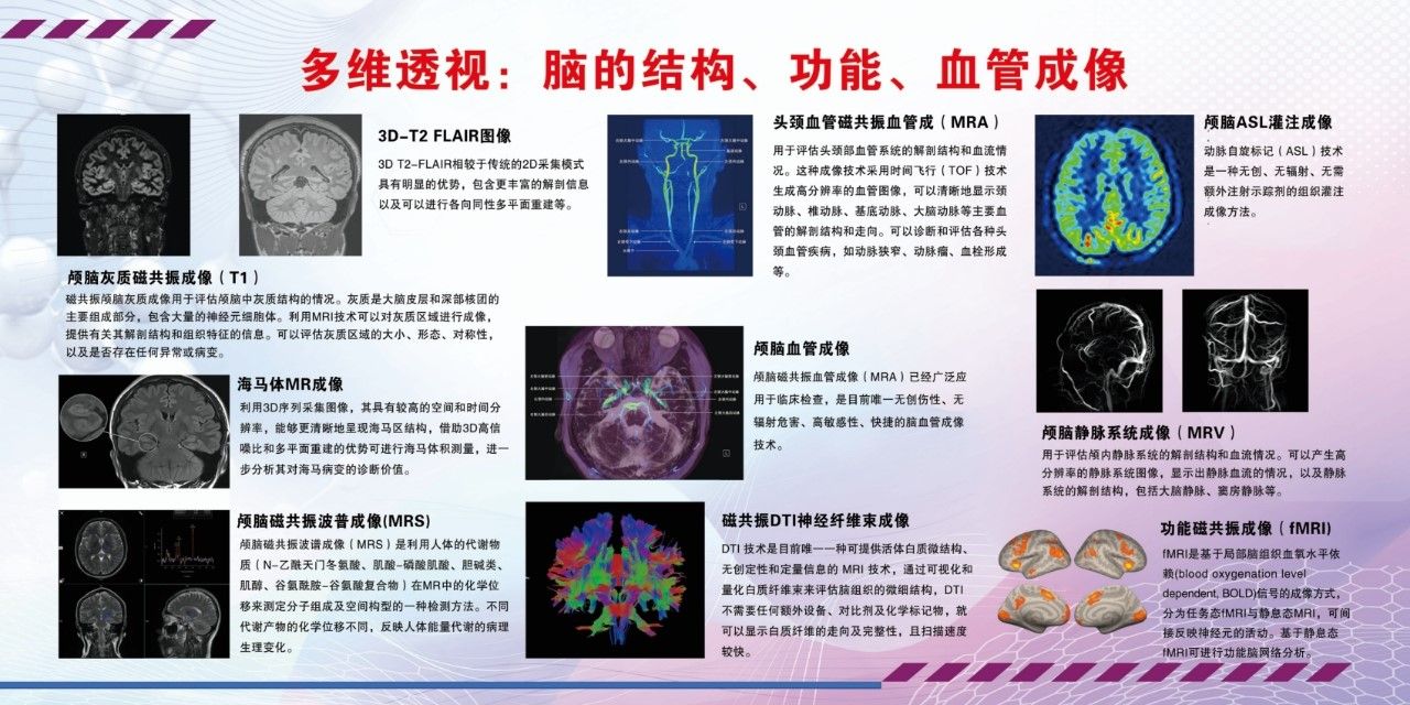
先进的成像设备就如科研工作者的“第三只眼”,可以帮助我们从各种层面和维度活体透视大脑,分析大脑结构、功能、血流、类淋巴等不同侧面的特征与规律(图2)。
As the "third eye" of scientists, the Siemens 3.0T Prisma system is helping the scientists to explore our brain in multi-levels and dimensions, analyze the characteristics and aspects of brain structure, function, blood flow, and lymph-like features (Figure 2).

图2多维透视:脑的结构、功能、血管成像
Figure 2 Multi-dimensional Perspective of Brain Structure, Function, and Vasculature
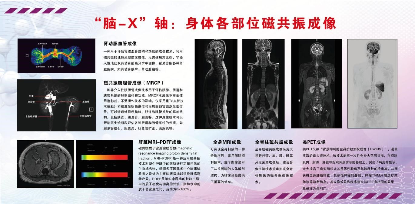
同时磁共振系统对于身体各个器官的成像技术日臻成熟,对于开展“脑-X”轴研究,探索中枢神经系统与躯体其他系统之间的密切联系大有帮助(图3)。
In addition, the MRI is also used to image various organs of the body, greatly aiding in the development of "brain-X" axis research, exploring the close connection between the central nervous system and other systems of the body (Figure 3).

图3 “脑-X”轴:身体各部位磁共振成像
Figure 3 "Brain-X" Axis: Magnetic Resonance Imaging of Various Parts of the Body
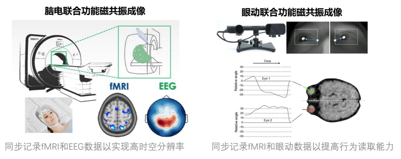
本中心依托西门子3.0T Prisma磁共振成像系统,通过与其它设备如Eyelink 1000 plus眼动仪、Brain Products (BP) 64导脑电采集系统等相结合,还可发挥多学科交叉、多模态数据融合的特色(图4)。
Combined with other devices such as the Eyelink 1000 plus eye tracker, Brain Products (BP) 64-channel EEG acquisition system, our MRI center can also perform the characteristics of interdisciplinary and the integration of multi-modal data (Figure 4).

图4 磁共振与脑电、眼动联采示意图
Figure 4 Schematic Diagram of MRI Combined with EEG and Eye Tracking Data Collection
中心工作环境宽敞明亮,配套设施齐全,配有专门的接待大厅、被试更衣室、洗衣房(用于换洗全棉被试服)、洗头间(用于脑电实验)等,也有专门的电子化神经心理评估室和独立的师生办公室、多媒体会议室等,方便课题组开展各类实验。
The center has a comfortable and efficient working environment with complete facilities, including a dedicated reception hall, subject changing room, laundry room (for changing cotton subject clothing), and a hair-washing room (for EEG experiments), as well as a dedicated electronic neuropsychological assessment room and independent offices for the researchers, and a multimedia conference room, making it convenient for research groups to carry out various studies.

图5 磁共振脑成像中心工作环境
Figure 5 Working Environment of the MRI Center
中心拥有专业的技术团队。目前拥有来自北京师范大学、北京邮电大学、山东省立医院、山东第一医科大学放射学院等单位的9名核心骨干教师、技师,并有2名在读博士研究生,3名在读硕士研究生,2名全职科研助理,以及若干名本科实习生,组成了一支技术过硬的服务队伍。本中心长期开放平台科研项目合作,并长期招募实习生与科研助理,欢迎有意向者与本中心联系,咨询邮箱:kxu@sdfmu.edu.cn(徐老师)。
The center is run by a professional technical team with nine core teachers and technicians from Beijing Normal University, Beijing University of Posts and Telecommunications, Shandong Provincial Hospital, and the School of Radiology of Shandong First Medical University, as well as two doctoral students, three master students, two full-time research assistants, and several undergraduate interns. The center is open to research project cooperation and is constantly recruiting interns and research assistants. We welcome those interested to contact our center for inquiries. The consultation email is kxu@sdfmu.edu.cn (Dr. Xu).
磁共振脑成像中心将秉持科学严谨的态度,结合先进技术和专业团队的力量,通过高质量、规范的成像和丰富的研究手段,助力推动脑科学与类脑研究的不断进步,促进本校在脑科学领域不断取得突破性进展。
The MRI Center will uphold a scientific and rigorous attitude, combining advanced technology and the strength of a professional team, through high-quality, standardized imaging and rich research methods, to promote the continuous progress of brain science and brain-inspired research, and to promote breakthroughs in the field of brain science.